Hypothesis
During speech, tongue shapes are created as the weighted sum of a small number of basic shapes.
Experiment
We tested the hypothesis through data reduction and factor analysis experiments applied to a series of tongue shapes derived from 3D MR images of the tongue during vowel and consonant production.
Results
- PARAFAC Analysis of the Three dimensional tongue Shape
- Electromagnetic exposure safety of the Carstens Articulograph AG100
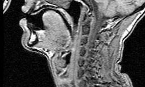
Biomedical Imaging of Speech Production
The Illinois Speech and Language Engineering group acquires and publishes biomedical imaging databases of speech production. The databases served from this web page were acquired in collaboration with the Illinois Speech and Hearing Sciences Department and the Illinois Biomedical Magnetic Resonance Facility. Image data currently available on this site include:
- The Vowel MRI database, collected at Cedars-Sinai Medical Center and segmented at UCLA while Professor Hasegawa-Johnson was a post-doc at UCLA, includes coronal and axial image stacks portraying five different speakers during production of the ten monophthongal vowels of English. One speaker also produces a selection of consonants. Tongue and palate are outlined in the coronal image stack only. Voice recordings were made shortly before or after the imaging session, in both prone and sitting positions in a quiet recording studio.
-
Microscopic
images of a 1cm cubic specimen of excised cadaver tongue
tissue. Tissue samples analyzed by Jerald Moon at the
University of Iowa, and by David Kuehn, Wei Tian, and Mark
Hasegawa-Johnson at the University of Illinois:
- Coronal MRI sections, pixel size 59×59 microns, slice thickness 49 microns, imaged by Victor Schepkin at the University of Illinois BMRF.
- Histologic sections of the same specimen, mounted at the University of Iowa.